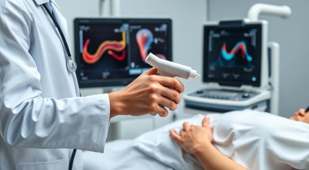
Renal, Portal, or Aorto-Iliac Duplex
Ultrasound has recently become an indispensable tool for family physicians, whether in primary care or emergency settings. It has also expanded into many other specialties such as internal medicine, intensive care, neurology, pulmonology, gastroenterology, etc., since ultrasounds have proven to be a safe tool with high diagnostic capacity. It is widely believed that bedside ultrasound can significantly complement the physical exam and greatly enhance a clinician's effectiveness, allowing the examiner immediate insight into the anatomy and physiology of certain structures. In this context, ultrasound studies of the aorta and major abdominal vessels have special relevance.
Renal Duplex
Renal artery Doppler ultrasound involves visualizing these arteries from the anterior abdominal wall. The images allow the arteries to be assessed and the blood flow velocity in the aorta and renal arteries to be measured. This way, physicians can confirm good blood supply to both kidneys, assess for possible stenosis or narrowing of any artery (which could cause treatment-resistant hypertension), verify proper function of prior interventions on renal arteries (bypass, stents, angioplasties), and rule out perfusion deficits that could result in kidney dysfunction of unknown origin.
This test is also performed to assess the blood vessels supplying the liver and nearby organs. It is generally used in patients with chronic liver disease or hepatitis to detect vascular complications. Doppler ultrasound is a precise method used to evaluate blood flow in the portal venous system, helping diagnose portal hypertension, assess the presence of portal-systemic shunts in more advanced cases, and guide procedures like transjugular intrahepatic portosystemic shunt (TIPS). It is also used in managing patients with gastrointestinal bleeding from various causes, among other indications.
Aorto-Iliac Duplex
This is an ultrasound study of the abdominal aorta. It involves visualization from the diaphragm down to the bifurcation of the common iliac arteries, using two-dimensional, color, and spectral Doppler imaging. As this study is performed on the abdominal cavity, a 6-hour fasting period is required for proper visualization of the aortic artery.
The main indications include screening for Abdominal Aortic Aneurysm, ruling out iliac aneurysm, aorto-iliac stenosis (Leriche’s Syndrome), and abdominal aortic dissection.
You can access this service through your insurance plan or as a private patient. Book your appointment and let us care for you — from heart to heart — with the support of our team of professionals.